
By Christian Espana Schmidt MD

This is a patient who comes with shortness of breath.
You obtain a heart ultrasound, can you tell me if the ejection fraction is normal or not?
The ejection fraction is abnormal. Look at the anterior leaflet of the mitral valve!

Is the EPSS normal?
One can quicly estimate the EPSS from here, this one specifically is more than 1.5cm
Other clues: Look at the ventricular cavity, it is not decreasing in size and the ventricular walls are not increasing its width as expected.

This is a "shallow" PLAX, from the same patient.
Any clue of what may be associated with shortness of breath?
Hoover in the answer below
If you guessed it is a pleural effusion you are right.
Behind the heart there is a large pleural effusion.
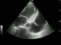
This is an apycal view with a twist
The left ventricle is not contracting and the mitral valve is not contracting normally. The left atria is enlarged and there is a large left pleural effusion.

This is the subcostal view of a heart with left ventricular failure, left atrial enlargement and you still can see the pleural effusion

This is an IVC with a blunted respirophasic response. Check the hepatic vein! It is also dilated.

Large right pleural effusion
At this moment I would ask you to listen to my video of lung ultrasound. Lern what is the curtain sign and where is this image is being obtained.

Large left pleural effusion
The difference between this one and the prior one is that the spleen is smaller and without the obvious vascular structures.
